
Phone:
+86-139-5121-3963
Views: 0 Author: Site Editor Publish Time: 2026-07-20 Origin: Site


The transition from open to laparoscopic liver resection hinges on overcoming the technical limitations of working in a confined space. Specifically, managing parenchymal transection and hemostasis presents a massive clinical hurdle. Uncontrolled bleeding and biliary leaks remain the primary reasons for conversion to open surgery. Advanced energy devices have become mandatory in the modern operating room, not just optional upgrades. Surgeons need reliable tools to navigate these high-stakes environments safely. Minor venous bleeding can instantly obscure the camera lens and ruin the surgical field of view.
An optimized ultrasound scalpel provides a precise balance of cutting and coagulation. It significantly reduces your reliance on multiple instruments during complex procedures. However, clinical evaluation requires careful analysis of lateral thermal spread, vessel sealing capabilities, and integration requirements. You will learn how these systems function mechanically, how they compare to traditional modalities, and what to consider for effective adoption in your facility.
Clinical Efficacy: Ultrasound activated scalpel systems simultaneously cut and coagulate, effectively managing vessels up to 5mm and reducing intraoperative blood loss.
Operational Efficiency: Decreases the need for constant instrument exchanges in the laparoscopic port, shortening total operative time.
Risk Mitigation: Delivers significantly lower lateral thermal spread compared to standard electrosurgery, protecting adjacent healthy liver parenchyma and biliary structures.
Procurement Reality: Successful adoption requires assessing the compatibility of the ultrasonic generator, calculating per-procedure consumable costs, and addressing the surgeon's learning curve.
We face severe surgical constraints when operating laparoscopically. You lose critical tactile feedback almost entirely. The surgical ports create restricted angles for instrument manipulation. These physical limitations make manual tying and suturing deeply inefficient during liver resections. Surgeons need faster, safer methods to secure tissue without fumbling with knots in a confined space.
The hemostasis imperative cannot be overstated. The liver features immense vascularity by nature. Even minor venous bleeding creates immediate problems. Blood absorbs light and obscures the camera lens instantly. You lose your field of view, forcing a halt to the procedure. Rapid, reliable hemostasis is not just a preference; it ensures patient safety and prevents emergency conversions to open surgery.
Biliary control represents another major hurdle. You must adequately seal small biliary radicles during parenchymal transection. Failure to achieve a tight seal leads to postoperative bile leaks. These leaks drive up morbidity rates significantly. They prolong hospital stays and increase patient discomfort. Proper energy application directly targets and prevents these severe complications.
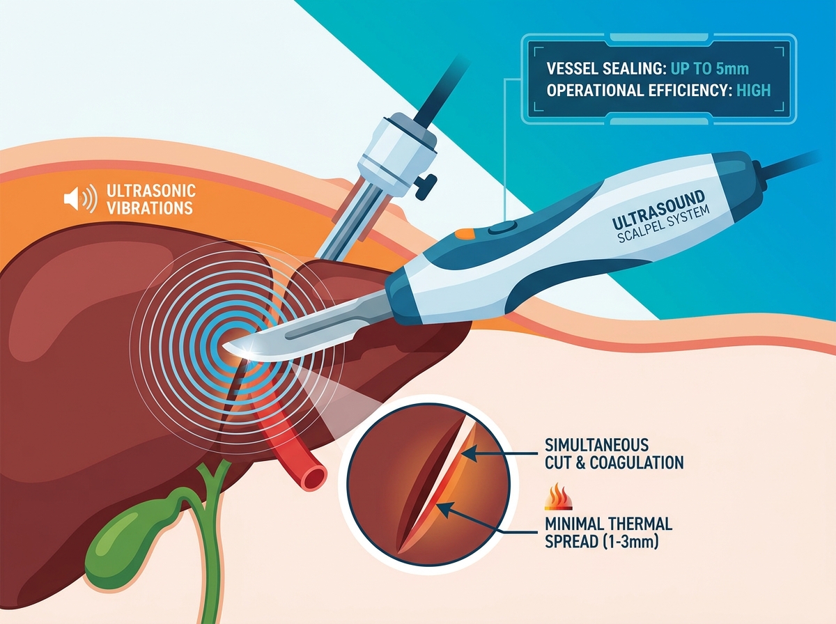
Surgeons often ask how these devices differ from traditional electrocautery. They use mechanical energy instead of electrical energy. An ultrasound activated scalpel system relies on high-frequency mechanical vibration. The blade typically oscillates at around 55.5 kHz. The system does not pass dangerous electrical current through the patient. This structural difference fundamentally protects vulnerable surrounding tissues.
The tissue effect relies on pure mechanical friction. Rapid vibration causes intracellular water to vaporize. It induces rapid protein denaturation within the vessel walls. This physical process forms a sticky, secure coagulum. The coagulum seals vessels perfectly before the blade divides them. You get a clean cut and a secure seal simultaneously without sparking.
The power source drives this entire process smoothly. An advanced ultrasonic generator translates electrical energy into piezoelectric motion in the handpiece. The generator plays a crucial role in surgical flow. It must maintain a stable resonant frequency. Tissue density changes constantly during dissection. Fibrotic or cirrhotic livers strongly resist blade movement. A quality generator adapts to these load changes instantly, maintaining cutting power.
You must evaluate any energy device against strict clinical metrics. We look at four primary dimensions during a laparoscopic liver resection to ensure safety and efficiency.
A laparoscopic ultrasonic scalpel must consistently seal vessels and ducts. Most quality devices handle diameters up to 3-5mm without requiring secondary surgical clips. This capability drastically reduces intraoperative blood loss. Historical data shows significant drops in blood transfusion rates when surgeons adopt ultrasonic energy over basic monopolar cautery.
Lateral thermal damage ruins healthy tissue. Ultrasonic energy stays highly localized by design. We typically observe a minimal lateral spread of 1-3mm. This precision minimizes necrosis of the remaining liver remnant. It critically protects adjacent major vessels and delicate biliary structures from delayed thermal breakdown.
The rapidly vibrating blade creates a cavitation effect in tissue fluids. This phenomenon helps separate loose parenchymal tissue safely. It exposes larger vessels cleanly without injuring them. You can then apply targeted clipping to these major structures. It functions beautifully as a blunt dissector.
Visibility governs everything in laparoscopy. Ultrasonic friction produces water vapor, not thick char smoke. This means significantly lower smoke production compared to standard electrocautery. You avoid endless camera cleaning interruptions and maintain procedural momentum.
Let's review these clinical metrics in a summary breakdown:
| Clinical Metric | Ultrasonic Performance Profile | Impact on Surgery |
|---|---|---|
| Vessel Sealing | Seals up to 5mm safely | Eliminates frequent clip application |
| Thermal Spread | 1-3mm lateral damage | Protects healthy liver remnant |
| Smoke Output | Minimal (mostly water vapor) | Maintains clear visual field |
| Cavitation | Separates loose parenchyma | Exposes major vessels cleanly |
Surgeons have several options for dividing tissue. Each modality carries specific strengths and inherent risks you must understand.
Standard electrosurgery effectively handles surface hemostasis. However, it generates high lateral thermal spread. It also produces excessive surgical smoke. These factors make it risky near major hepatic veins. You risk thermal injury to vital structures, leading to catastrophic postoperative bleeding.
CUSA remains the gold standard for skeletonizing vessels in open surgery. It selectively destroys parenchyma while sparing vessels. Yet, it proves cumbersome in purely laparoscopic setups. It operates slower in tight spaces. It also requires secondary instruments to actually seal and cut the exposed vessels, adding steps to the procedure.
Ultrasonic energy blends multiple functions into one cohesive device. It acts simultaneously as a dissector, grasper, coagulator, and cutter. This multifunctionality dramatically reduces the "instrument exchange" bottleneck inherent to laparoscopy. Every time you swap tools in a laparoscopic port, you lose valuable operative time. Consolidating functions keeps the operation moving smoothly and reduces surgeon fatigue.
Deploying these advanced devices requires careful planning. You cannot simply hand them to surgeons without protocol training. Several practical realities govern successful adoption.
First, we must acknowledge the surgeon learning curve. Device mishandling leads to failure. "Blade fatigue" occurs frequently if misused. Tip breakage happens if surgeons apply excessive torque during dissection. Activating the vibrating blade directly against metal clips will shatter the tip instantly. Proper education prevents these expensive and dangerous errors.
Managing tissue tension dictates success. Ultrasonic devices require specific traction and counter-traction techniques. The blade needs proper resistance to function optimally. Improper tension leads to incomplete seals. If the tissue is too loose, it won't cut cleanly. If it is pulled too tight, the vessel tears before it seals.
Financial evaluation centers on capital versus consumable expenditures. You must balance the upfront capital expenditure of the ultrasonic generator. You then weigh this against the recurring costs of single-use handpieces per procedure. Accurate budget forecasting ensures sustainable access to the technology.
Infrastructure compatibility matters heavily in crowded operating rooms. Evaluate the physical footprint in your OR tower. Determine whether universal generators can power both your ultrasonic and advanced bipolar devices. Reducing tower clutter improves nursing efficiency and saves floor space.
Hospitals must employ structured evaluation processes. Value Analysis Committees face tough decisions when approving new surgical technology.
To justify the capital spend, deeply evaluate standardization potential. Can this platform be used across general surgery, gynecology, and urology? A versatile generator provides much higher utility for the hospital. It prevents the need to buy separate systems for different surgical departments.
Demand evidence-based evaluation. Do not rely solely on sales marketing claims. Request peer-reviewed clinical trial data. The data must be specific to major and minor hepatectomies. Liver tissue behaves differently than bowel or muscular tissue, so context matters immensely.
Trial structuring determines accurate feedback. Set clear clinical success criteria before bringing the device into the OR. Consider tracking these exact metrics during hands-on evaluations:
Total parenchymal transection time per case.
Estimated intraoperative blood loss.
Frequency of instrument exchanges.
Instances of device failures or blade breakages.
An ultrasound scalpel is not a magic bullet. It does not replace sound surgical technique or deep anatomical knowledge. However, it remains a proven necessity for safe, efficient laparoscopic liver resections. It bridges the gap between precision and speed in a highly restrictive environment.
For your next steps:
Prioritize verifiable thermal spread data during technical reviews.
Assess generator reliability and cross-specialty compatibility to maximize utility.
Implement mandatory training on proper tissue tension to prevent tip breakages.
Monitor early clinical outcomes to validate your procurement choice.
A: Most commercial systems are FDA-cleared to seal vessels up to 5mm in diameter. Larger hepatic veins still require endo-staplers or polymer clips.
A: Typically, yes. Most manufacturers engineer a closed-loop system where the generator and handpiece communicate to monitor tissue impedance and blade temperature.
A: Ultrasonic devices generally exhibit a smaller lateral thermal spread (1-3mm) compared to standard bipolar, though advanced bipolar systems have narrowed this gap. The choice often depends on whether dissection speed (ultrasonic) or large-vessel sealing capability (bipolar) is prioritized.


