
Phone:
+86-139-5121-3963
Views: 0 Author: Site Editor Publish Time: 2026-09-14 Origin: Site


When surgical procurement teams or clinical boards ask about the diameter of a vessel sealer, they often face a critical ambiguity. Are they evaluating the maximum vessel diameter the device can safely fuse? Or are they questioning the instrument shaft diameter required for trocar insertion? Both metrics heavily influence success in the operating room. Selecting the right device requires balancing maximum sealing capability alongside minimally invasive access requirements. You must strictly adhere to safety clearances to prevent postoperative hemorrhage. Misunderstanding these specifications risks patient safety. It also disrupts standard surgical workflows. Our goal here is to provide a technical breakdown of these vital dimensions. We evaluate current industry standards to clarify the confusion. You will learn how to build a clear, actionable decision framework for hospital procurement and surgical teams.
Vessel Sealing Capacity: The industry standard and regulatory (e.g., FDA) clearance for advanced bipolar and ultrasonic vessel sealers is typically up to and including 7 mm in diameter for arteries and veins.
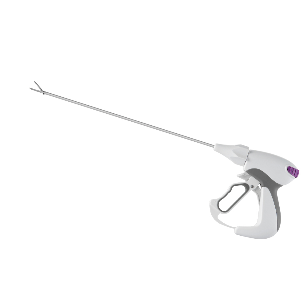
Instrument Shaft Size: A modern laparoscopic vessel sealing device generally features a 5 mm shaft diameter to optimize minimally invasive access, though legacy or specialized 10 mm devices exist.
Evaluation Criteria: Procurement decisions should prioritize not just the 7mm claim, but consistent burst pressure (typically 3x normal systolic pressure), minimal thermal spread, and jaw design compatibility with specific surgical specialties.
Risk Mitigation: Relying on a vessel sealer for vessels exceeding 7 mm or heavily calcified vessels poses severe clinical risks; back-up modalities (clips, staplers) must be part of the surgical plan.
Device specifications often confuse new procurement staff. You must clearly separate the two distinct dimensions evaluated during shortlisting. Mixing them up leads to purchasing errors. We categorize these metrics into clinical indications and hardware footprints.
When you read a product brochure, you will see two numbers. You must map each number to its specific function in the operating room.
Vessel Diameter (Clinical Indication): This represents the anatomical size limit. It measures the tissue bundle, artery, or vein the device can safely fuse. Standard clearances usually peak at 7 millimeters. This limit dictates surgical safety. It ensures the tissue receives adequate energy to form a permanent collagen seal.
Shaft Diameter (Hardware Footprint): This measures the physical thickness of the device shaft. It dictates which surgical trocars or ports you must use. The outer tube houses the internal mechanisms. Surgeons rely on this dimension to plan patient incisions. Smaller shafts mean less invasive entry points.
Misalignment between these two metrics causes cascading problems. If a device shaft size does not match standard hospital trocar inventory, you face severe OR delays. Surgical teams will open sterile packaging only to realize the tool cannot fit the port. This mismatch wastes single-use items. It forces staff to scramble for alternative instruments.
Furthermore, misunderstanding vessel capacity leads to severe clinical adverse events. If a surgeon assumes a device can handle an 8mm artery because the shaft is 10mm wide, catastrophic bleeding may occur. The hardware footprint never dictates sealing capacity. You must evaluate both numbers independently. Clear communication between supply chain and clinical staff prevents these dangerous assumptions.
Dimension Specifications and Clinical Impact Summary
| Dimension Type | Standard Measurement | Clinical Focus | Procurement Impact |
|---|---|---|---|
| Vessel Diameter | Up to 7 mm | Tissue fusion, hemostasis, safety | Determines surgical indications and specialty fit |
| Shaft Diameter | Typically 5 mm | Incision size, trocar compatibility | Drives inventory standardization and port selection |
The 7mm threshold represents a cornerstone of modern surgical safety. Devices undergo rigorous testing to prove they can reliably fuse vessels of this size. Understanding how this process works builds confidence in the operating room.
Advanced bipolar and ultrasonic technologies utilize a combination of mechanical compression and energy delivery. When surgeons clamp the jaws, the instrument applies precise pressure to the vessel walls. The generator then delivers energy into the tissue.
This energy denatures collagen and elastin proteins within the vessel structure. As the proteins melt, they intermingle and reform. They create a translucent, permanent plastic-like seal. This seal withstands significant internal pressure. Fusing a full 7mm vessel requires highly calibrated energy algorithms. The system must measure tissue impedance constantly. It stops the energy flow exactly when the fusion completes.
Regulatory bodies enforce strict testing protocols. Standard FDA-cleared indications cover vessels up to 7 mm. Manufacturers must submit extensive data to achieve this clearance. They cannot simply claim the device works on large arteries.
The core safety benchmark involves burst pressure testing. A reliable seal must withstand at least three times the normal systolic blood pressure. Healthy human systolic pressure peaks around 120 mmHg. Therefore, regulators require seals to withstand 360 mmHg or higher. Top-tier devices often demonstrate burst pressures exceeding 500 mmHg. This buffer guarantees the seal holds even if the patient experiences postoperative hypertension.
Anatomical structures differ significantly. While devices hold clearances for 7mm arteries and veins, surgeons encounter various tissue types. Tissue bundles, broad ligaments, and lymphatic vessels are routinely sealed. However, their structural composition differs from distinct vascular structures.
Veins have thin walls. Arteries have thick, muscular walls. Lymphatics carry fluid but lack robust collagen structures. A 7mm artery presents a different challenge than a 7mm vein. The device algorithm must adapt to these physical differences automatically. It adjusts energy output based on the specific resistance of the clamped tissue.
You must maintain a healthy skepticism during device evaluation. Warn your teams against assuming all devices perform equally on a 7mm vessel. Two tools might both possess FDA clearance for 7mm arteries. However, variations in energy delivery algorithms deeply affect the speed and reliability of the seal.
Some devices seal a 7mm vessel in three seconds. Others might take seven seconds and generate excessive smoke. Some require multiple overlapping bites. Slower algorithms increase surgeon fatigue. They also prolong the total operation time. Always demand comparative performance data rather than accepting baseline regulatory claims.
The physical footprint of a laparoscopic vessel sealing device dictates surgical access. Surgeons constantly strive for less invasive approaches. Instrument design has evolved to meet these demands without sacrificing jaw power.
Today, 5mm is the dominant shaft diameter for modern laparoscopic instruments. This standard emerged for several compelling clinical reasons. Primarily, it minimizes patient incision size. Smaller incisions directly correlate to reduced postoperative pain. They lower the risk of port-site hernias. They also improve cosmetic outcomes.
A 5mm shaft allows tremendous versatility across port sizes. You can insert it directly into a standard 5mm trocar. You can use it in an 8mm robotic port. You can also pass it through a 12mm port using a simple reducer cap. This flexibility makes the 5mm standard universally acceptable across various surgical disciplines.
A fascinating engineering challenge exists within these tools. You must contrast the narrow shaft diameter with the required jaw length and opening angle. A 5mm shaft must still deploy a jaw wide enough to capture a 7mm vessel. It also needs enough structural strength to apply high compression forces.
Achieving this requires sophisticated hinge mechanisms. Engineers design double-action jaws maximizing the opening angle. They incorporate strong, miniature blade tracks for tissue transection. The jaw must taper at the tip for fine dissection. Yet, the base must remain robust enough to hold thick tissue bundles. The transition from a slender 5mm tube to a powerful crushing jaw represents a peak of medical engineering.
Standardizing to 5mm devices deeply streamlines inventory management. Hospital supply rooms often overflow with redundant items. By committing to the 5mm standard, you reduce the need for multiple trocar sizes in the OR.
Surgical techs can build standard, universal laparoscopy trays. They do not need to pack separate 10mm ports just to accommodate one bulky legacy sealer. This standardization accelerates OR turnover times. It simplifies the setup process. It removes guesswork during emergent procedures.
Dimensions represent only the baseline. You must evaluate features directly translating to surgical outcomes. When finalizing a hospital shortlist, look beyond basic diameter measurements.
Clinical performance hinges on several secondary characteristics. You need to scrutinize how the instrument interacts with surrounding anatomy.
Thermal Spread: This measures how far heat travels beyond the jaws. You typically want thermal spread targeting under 2mm. This is crucial for operating near vital structures. When working near the recurrent laryngeal nerve or the ureter, excessive heat causes devastating permanent damage.
Cutting Mechanism: Devices either feature an independent manual blade or integrated energy-based cutting. Manual blades give surgeons tactile control over transection. Energy-based cutting removes the physical blade. It relies entirely on targeted heat to divide the tissue. You should base this choice on specific surgical specialty preferences.
Ergonomics and Haptics: Long surgeries cause hand cramps. Surgeon fatigue reduction relies heavily on handle design. An intuitive grip angle minimizes wrist strain. Furthermore, audible feedback remains essential. The generator must provide clear audio tones indicating the seal cycle is complete or if an error occurred.
Procurement teams must carefully evaluate the ecosystem of the devices. Assess whether you need single-use disposable devices or reusable devices featuring disposable tips. Reusable handles reduce medical waste. They require robust sterilization workflows.
You must factor in compatibility. Many advanced sealers only function when plugged into their proprietary electrosurgical generators. If you select a new brand of sealer, you might need to acquire an entirely new fleet of capital equipment. Evaluate your current generator fleet before shortlisting a new instrument brand. Streamlining generator platforms simplifies OR infrastructure.
Even the most advanced instrument poses risks if used improperly. Clinical teams must establish rigorous protocols. Training bridges the gap between device capabilities and actual patient safety.
Vessels exceeding 7mm require strict management protocols. You must establish clear rules regarding anatomical limits. If a vessel appears larger than 7mm, energy alone is insufficient. Similarly, highly calcified or atherosclerotic vessels resist proper compression. The energy cannot denature the diseased collagen effectively.
In these scenarios, you must secure the vessel using mechanical methods. Surgeons must apply mechanical clips, traditional sutures, or vascular staplers. Relying on an energy seal for a calcified 9mm artery invites disastrous postoperative hemorrhage. Back-up modalities must always be available on the sterile field.
Transitioning to a new vessel sealer requires tactile adjustment. Every device feels different. The activation button placement varies. The required grip force changes. The tissue bite sizes differ.
Surgeons must undergo dedicated training. They must learn not to prematurely interrupt the automated energy cycle. If a surgeon opens the jaws before the generator sounds the completion tone, the seal remains weak. They must trust the impedance feedback loop. Patience during the three-to-five second cycle guarantees a permanent tissue fusion.
Technology does not replace clinical judgment. We emphasize that despite device claims, surgeons must always visually verify the seal. Before activating the cutting blade, the surgeon must inspect the tissue.
Look for translucency: A proper seal looks somewhat clear, like plastic.
Check for blanching: The surrounding tissue should show a distinct change in color.
Assess for uniform compression: The flattened area should appear consistent across the entire jaw width.
If the seal looks incomplete or charred, the surgeon must reapply the instrument. Never blindly transect tissue without this critical visual confirmation.
The "diameter" question defines the fundamental baseline for device selection. You now know it implies two distinct metrics. It demands 7mm clinical sealing capability paired seamlessly alongside a 5mm laparoscopic footprint. Aligning these numbers ensures surgical safety and operational efficiency.
Procurement teams should take immediate action. First, request in-service clinical trials to test ergonomics and thermal spread. Next, review independent burst-pressure data rather than relying solely on marketing brochures. Finally, rigorously assess generator compatibility across your facility. Addressing these steps guarantees a safer operating room and a smoother supply chain.
A: No. Using energy devices on vessels exceeding their FDA-cleared diameter (typically 7mm) increases the risk of seal failure and hemorrhage. Mechanical methods are required. You must utilize sutures, clips, or vascular staplers for larger or highly calcified arteries.
A: Both top-tier modalities are generally cleared for up to 7mm vessels, but they achieve the seal differently. Bipolar relies on tissue impedance feedback, while ultrasonic relies on mechanical friction. Surgeon preference and specific thermal spread requirements usually dictate the choice.
A: While shaft diameter is typically 5mm, the working length usually ranges from 37cm to 44cm. This variation accommodates different patient BMIs and surgical approaches. For example, bariatric procedures require longer shafts than standard pelvic surgeries.


