
Phone:
+86-139-5121-3963
Views: 0 Author: Site Editor Publish Time: 2026-09-29 Origin: Site


Safe tissue extraction is a critical operational imperative during laparoscopic procedures such as cholecystectomies, appendectomies, and oncology resections. Modern surgical teams rely on precise instrumentation to isolate and remove excised tissues without compromising the sterile field. However, improper specimen handling introduces severe clinical risks. Failing to secure the tissue properly often leads to devastating port-site metastasis, unintended tissue fragmentation, and dangerous surgical site infections (SSIs). These avoidable complications jeopardize patient safety while significantly straining hospital resources. This guide provides an evidence-oriented framework for both surgical teams and supply chain managers. You will discover practical steps for proper deployment and effective strategies to evaluate device efficacy. Furthermore, we will show you how to standardize your retrieval protocols to ensure consistent, safe outcomes in every minimally invasive case.
Successful use of an endo catch bag relies on precise trocar sizing, controlled deployment, and strict adherence to extraction protocols to prevent bag rupture.
Evaluating a specimen retrieval bag requires analyzing material tensile strength, deployment mechanisms (automatic vs. manual), and volume capacity relative to procedural volume.
Standardizing high-quality retrieval devices reduces operative time, minimizes cross-contamination risks, and supports compliance with stringent infection control mandates.
Cross-disciplinary input (from surgeons to supply chain) is critical when shortlisting devices for facility-wide adoption.
Defining a successful tissue extraction requires looking beyond simply removing the organ. Clinical success criteria demand zero spillage of contaminated fluids. Pathologists require intact specimen margins to conduct accurate histological evaluations. Furthermore, the patient needs minimal tissue trauma at the extraction site. Achieving these goals consistently requires standardized, purpose-built surgical instruments.
Surgical teams traditionally relied on open grasping techniques or makeshift extraction methods. We now recognize these older methods fall short of modern clinical standards. Today, closed-pouch systems stand as the definitive solution for mitigating tumor seeding and bacterial contamination. An endo catch bag encapsulates the excised mass entirely before it passes through the abdominal wall. This complete encapsulation prevents malignant cells or purulent material from contacting the incision margins.
Navigating the procurement landscape requires understanding basic terminology alignment. Medical professionals often use terms interchangeably across different regions and facilities. You will frequently hear the terms endo retrieval bag and specimen retrieval bag used to describe the exact same clinical function. However, specific functional mechanisms differ heavily by manufacturer. Some models feature a memory-wire auto-opening system. Others utilize a manual push-pull deployment method. Understanding these mechanical nuances helps clinical teams select the most appropriate device for their specific surgical environments.
Surgical success begins before the device ever enters the patient. You must verify device sizing against the primary trocar. Manufacturers typically engineer these devices for specific 10mm or 15mm trocar requirements. Inserting a larger delivery shaft into an undersized trocar will damage the device housing. This forces unnecessary instrument exchanges and delays the procedure.
Best practices dictate preparing the surgical field meticulously. Ensure the camera angle provides clear, unobstructed visualization prior to introduction. Clear away any surrounding omentum or bowel loops. A well-prepared visual field allows the surgeon to introduce the device safely without causing inadvertent visceral injuries.
Execution requires smooth, deliberate movements. Follow these structured steps to ensure safe capture:
Insert the delivery shaft: Advance the instrument smoothly through the designated trocar until the tip reaches the target area.
Deploy the pouch: Activate the device using its specific mechanism. Many modern tools utilize an ergonomic thumb ring or a sliding plunger. Push the mechanism fully to deploy the pouch into the abdominal cavity.
Position the bag: Orient the open mouth of the bag toward the excised organ. If utilizing a memory-wire model, allow the ring to expand fully.
Maneuver the tissue: Utilize atraumatic graspers to handle the specimen. Gently place the excised tissue directly into the center of the open pouch. Avoid grasping the bag material directly to prevent tearing.
Confirm seating: Verify the specimen rests fully below the closure line. Tissues resting near the rim will pinch during the sealing process.
Once the specimen sits securely inside the pouch, you must close the system immediately. Engage the cinch or closure mechanism to seal the bag mouth tightly. A secure seal traps all fluids and prevents intra-abdominal spillage.
Next, detach the bag from the delivery shaft. Not all models allow detachment, so you must know your specific device design. For non-detachable models, withdraw the entire apparatus slowly through the fascial defect. Pulling too quickly increases friction and risks fascial trauma.
Large specimens present a unique challenge. You cannot pull a massive spleen or grossly distended gallbladder through a standard 10mm port. In these cases, you must extend the fascial incision. Keep the neck of the bag exteriorized. Alternatively, teams utilize manual morcellation inside the bag. Surgeons break down the tissue carefully using ring forceps while the bag protects the surrounding anatomy. This protocol prevents catastrophic bag ruptures during extraction.
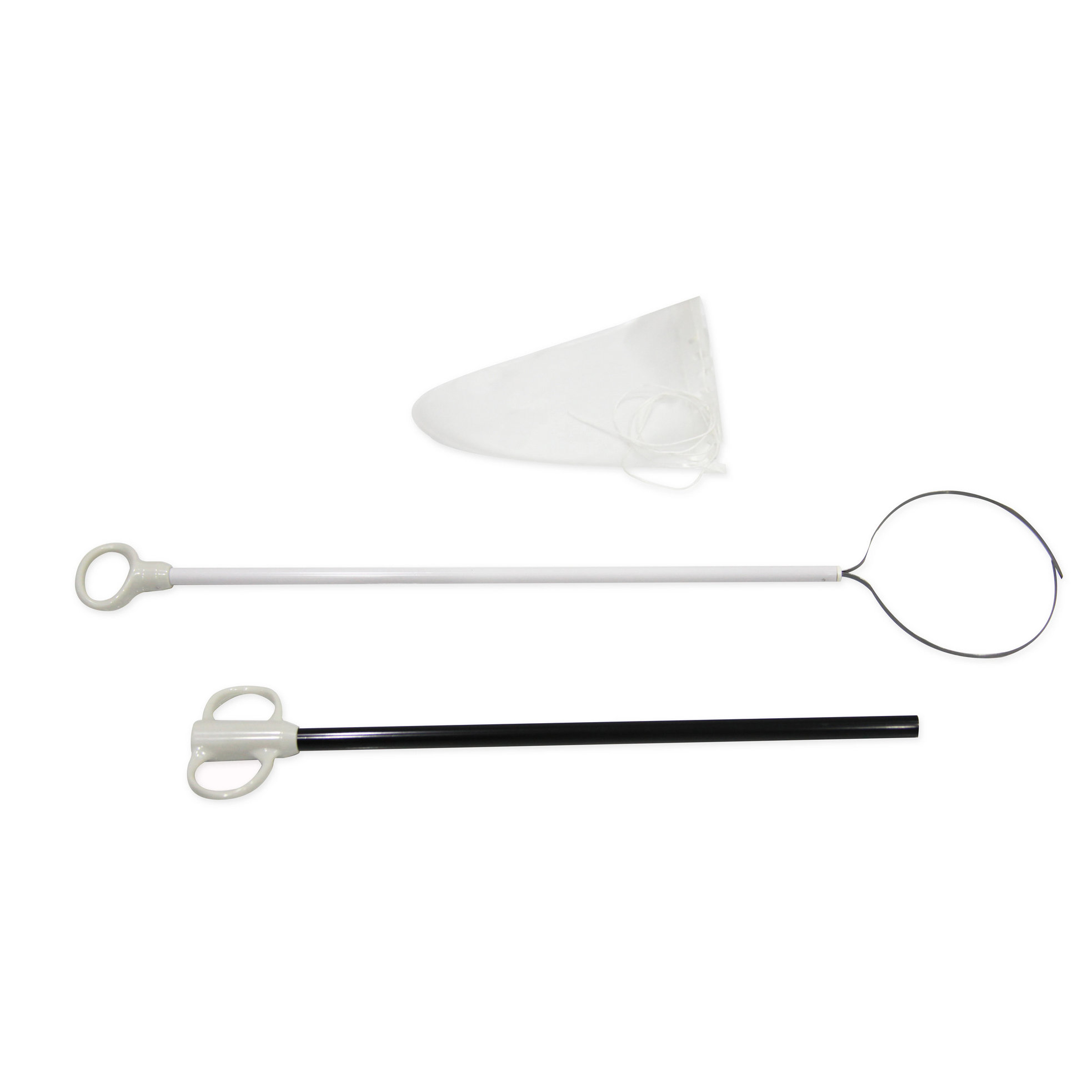
Even the most experienced surgical teams face material failure risks. Addressing the realities of material stress remains paramount for patient safety. High-quality devices utilize polyurethane bags because they offer exceptional durability and fluid retention. However, polyurethane has distinct tensile limits. Applying excessive force during a difficult extraction stretches the polymer structure. This extreme tension frequently leads to micro-perforations. Micro-perforations allow contaminated fluids to leak invisibly into the surgical site.
The intra-abdominal surgical environment presents additional hazards. The pneumoperitoneum acts as a busy workspace filled with hazardous tools. Consider the following environmental risks:
Sharp instrument proximity: Scissors and scalpel blades operating near the bag can inadvertently slice the polyurethane barrier.
Electrocautery damage: Active energy devices emit intense heat. Even brief contact between an active hook and the plastic pouch causes immediate melting and structural failure.
Suture snags: Stray needles or rough fascial edges can catch the bag material during the final extraction phase.
Mitigating these risks requires strict procedural discipline. We strongly recommend standardizing visual inspections post-extraction. Once you remove the bag, examine it under direct light. Confirm zero leakage and verify structural integrity. If you suspect any breach occurred during the procedure, initiate secondary wash-out protocols immediately. Flushing the surgical site reduces the bioburden and mitigates potential infectious complications.
Procurement teams must evaluate core material specifications rigorously. You should prioritize tensile strength, tear resistance, and overall impermeability. Do not rely on marketing claims alone. Look for evidence-backed testing data regarding fluid retention under pressure. A reliable device must maintain absolute integrity when subjected to extreme intra-abdominal forces.
The opening mechanism dictates usability. Surgeons prefer devices offering seamless single-handed operation. Memory wire systems self-open upon deployment, creating a wide, stable target for the tissue. String-pull mechanisms require more manual manipulation but often fit through smaller port sites. Review the chart below to compare common deployment styles.
Chart: Deployment Mechanism Comparison
| Mechanism Type | Deployment Method | Primary Clinical Advantage | Common Limitation |
|---|---|---|---|
| Memory Wire (Auto) | Spring-loaded release via plunger | Maintains open shape instantly | Requires larger trocar diameter |
| Manual String-Pull | Push rod and manual string tension | Fits through 5mm/10mm ports easily | Requires two-handed operation |
| Detachable Pouch | Bag releases from delivery shaft | Allows multiple instrument uses | Re-grasping the bag can be difficult |
A single bag size cannot accommodate every surgical discipline. Hospital inventory must match the anticipated clinical case mix. Using an oversized bag for a tiny specimen wastes resources and clutters the visual field. Conversely, using an undersized bag for a large organ guarantees structural failure.
Proper inventory management involves stocking various capacities. Small bags serve well for appendectomies and ectopic pregnancies. Medium bags handle routine cholecystectomies safely. High-volume bags remain necessary for major oncology resections, splenectomies, and complex nephrectomies. Veterinary clinics also require specialized sizes to accommodate diverse animal anatomies.
Table 1: Volume Capacity and Clinical Use Cases
| Size Category | Typical Volume (mL) | Target Procedures |
|---|---|---|
| Small | 100mL - 200mL | Appendectomy, Ovarian Cysts |
| Medium | 200mL - 400mL | Cholecystectomy, Lymph Node Biopsy |
| Large / High-Volume | 500mL - 1500mL+ | Splenectomy, Nephrectomy, Bowel Resection |
Patient safety hinges on strict regulatory compliance. Ensure every selected device meets rigorous FDA or CE mark standards. These certifications guarantee the materials pass strict biocompatibility and sterilization testing. Devices lacking proper certification introduce severe legal and clinical liabilities. Specialized facilities might also require additional certifications for advanced robotic platforms. Always request official compliance documentation during the vendor evaluation phase.
Implementing a new surgical device facility-wide presents unique logistical challenges. Supply chain managers naturally desire vendor consolidation. Single-sourcing devices streamlines ordering processes and reduces inventory complexity. However, surgical demand often pushes back against consolidation. Surgeons require procedure-specific pouch designs to optimize their distinct workflows. Balancing these competing interests requires open, cross-disciplinary communication.
We recommend establishing an evaluation committee comprising lead surgeons, operating room directors, and procurement specialists. This committee can review clinical outcomes and operational needs holistically. They ensure the chosen product satisfies the financial strategy while meeting strict clinical demands.
Acknowledge that introducing any new device requires dedicated in-service training. Surgical technicians and circulating nurses must understand the deployment mechanics intimately. Smooth handoffs prevent intra-operative delays. Organize hands-on training sessions using simulated abdominal trainers. Allow the staff to practice deploying, sealing, and extracting the bag repeatedly. Familiarity breeds confidence, and confident teams execute procedures safely.
Mastering proper extraction techniques is as much about choosing a reliable, compliant device as it is about surgical technique. Operating room teams must balance precise handling skills alongside rigorous equipment evaluation. An effective protocol eliminates spillage, protects the patient from metastatic spread, and accelerates overall recovery times.
Take proactive steps today to elevate your surgical standards. We encourage clinical directors and procurement teams to request physical product samples from reputable manufacturers. Review the clinical outcome data thoroughly. Furthermore, schedule an in-service evaluation to test the device in a controlled, simulated environment. Empowering your surgical staff through hands-on evaluation guarantees safer procedures and vastly improved patient outcomes.
A: Standard bags cannot safely accommodate motorized morcellation. Power morcellators easily tear through standard polyurethane walls, risking massive tissue spillage. You must use specialized contained morcellation systems. These advanced systems feature heavily reinforced, puncture-resistant materials specifically engineered to withstand blade contact.
A: Surgeons systematically enlarge the fascial incision to accommodate the bulk. To maintain the sterile barrier, they carefully exteriorize the mouth of the bag through the incision. They then extract the tissue piecemeal or gently pull the intact bag through the widened fascial defect.
A: Yes. Manufacturers design these devices strictly for single-use applications. Attempting to reprocess or sterilize them degrades the polyurethane material. This degradation destroys the tensile strength and introduces severe cross-contamination risks to future patients.
A: The core deployment principles remain identical across disciplines. However, veterinary surgeons face drastically different anatomical spaces and organ proportions. Veterinary procedures often utilize specialized bag sizes and adapted port placements to accommodate the unique physiology of non-human patients.


