
Phone:
+86-139-5121-3963
Views: 0 Author: Site Editor Publish Time: 2026-07-27 Origin: Site


Establishing safe primary abdominal access remains the absolute most critical first step in laparoscopic cholecystectomy. Surgeons must actively minimize severe vascular and visceral injury during this initial phase. Historically, clinical teams relied heavily on traditional blind entry using a Veress needle. Others preferred the reliable but highly time-consuming open Hasson technique. Today, modern surgical facilities increasingly adopt advanced optical entry methods. They offer a fundamentally safer and highly predictable approach for patients.
Why do surgical specialists strongly prefer a 10mm port for primary umbilicus entry? This specific size perfectly balances immediate tissue-layer visualization against necessary instrument capacity. It accommodates vital gallbladder extraction tools effortlessly. By reading this comprehensive guide, you will understand the precise mechanics of visual entry. We will outline essential procurement evaluation criteria for your operating room. Finally, you will learn how to evaluate medical suppliers objectively to ensure optimal patient outcomes.
Safety through Visualization: An optical visualization trocar allows real-time, layer-by-layer optical feedback during insertion, significantly mitigating blind entry risks.
Procedural Efficiency: The 10mm sizing is universally compatible with standard laparoscopes, 10mm clip appliers, and extraction bags required for cholecystectomy workflows.
Evaluation Criteria: Procurement and surgical teams should prioritize ergonomic control, clear distal lens design, and robust seal integrity when shortlisting trocar vendors.
Primary port placement carries the highest risk of iatrogenic injury during any laparoscopic procedure. Surgeons face a blind approach when creating this initial access point. Misjudging insertion depth can fatally damage major retroperitoneal blood vessels. It can also puncture underlying bowel loops. Hospitals face major clinical setbacks and intense legal scrutiny when these entirely preventable complications occur.
We must rigorously compare current entry modalities to understand the shift in best practices. The traditional Veress needle relies entirely on tactile feedback. Clinicians literally wait to feel specific "pops" through tissue planes. This tactile-only approach leaves too much room for dangerous human error. It relies on subjective sensation rather than objective evidence. Conversely, the open Hasson technique offers excellent safety. However, it requires significant manual labor. Surgeons must manually incise the fascia, place stay sutures, and close the defect meticulously afterward. This process consumes valuable operating room time.
Modern surgical facilities desperately need a middle ground. They demand speed, uncompromising safety, and procedural reliability. Integrating a high-quality laparoscopic trocar adds immense value by solving these historical challenges.
To measure true access success, clinical teams utilize strict, non-negotiable criteria. We target the following key benchmarks for primary entry:
Zero insertion-related organ trauma: The chosen device must never puncture underlying viscera blindly.
Minimal fascial tearing: The entry method should gently part muscle fibers rather than severing them abruptly.
Immediate pneumoperitoneum: The port must establish and hold insufflation gas rapidly to expand the surgical workspace.
An optical trocar fundamentally revolutionizes how surgeons enter the human abdomen. You integrate a standard 0-degree laparoscope directly inside the hollow obturator before initiating insertion. The clear distal tip acts as an illuminated window into the patient's anatomy.
This simple mechanism delivers a profound clinical outcome. You gain crystal-clear identification of every single anatomical layer. You see the yellow subcutaneous fat initially. You watch the tough rectus fascia part around the tip. You identify the dense linea alba clearly. Finally, you view the thin, translucent peritoneum just before entering the peritoneal cavity. This real-time, high-definition feedback completely eliminates dangerous guesswork from the procedure.
Traditional older ports utilize sharp, metal bladed tips. They slice aggressively and permanently through abdominal wall muscles. Bladeless tissue dilation works entirely differently. It utilizes a tapered, symmetric geometry. It gently separates tissue fibers laterally as you apply downward pressure. It pushes delicate muscle fibers aside rather than amputating them.
The measurable clinical benefits of bladeless dilation are overwhelming. Patients experience significantly reduced fascial defects upon device removal. Consequently, they face dramatically lower postoperative incisional hernia rates. Furthermore, the laterally dilated tissue naturally grips the ribbed cannula tightly. This dynamic creates much firmer port fixation at the umbilicus. You will experience far less frustrating slippage during rapid instrument exchanges.
A well-manufactured 10mm optical trocar aligns flawlessly with standardized surgical workflows. First, it easily accommodates standard 10mm optics. This provides the primary high-definition, wide-angle view required for safe cystic duct dissection.
Secondly, this specific sizing drastically boosts final specimen extraction efficiency. Intact gallbladder removal requires very specific tools. You need immediate passage for 10mm polymer clip appliers to secure the cystic duct and artery safely. Later, you must deploy a reliable extraction bag into the cavity. A 10mm port facilitates this intact gallbladder removal seamlessly. You do not need to pause the surgery to painfully enlarge the port site.
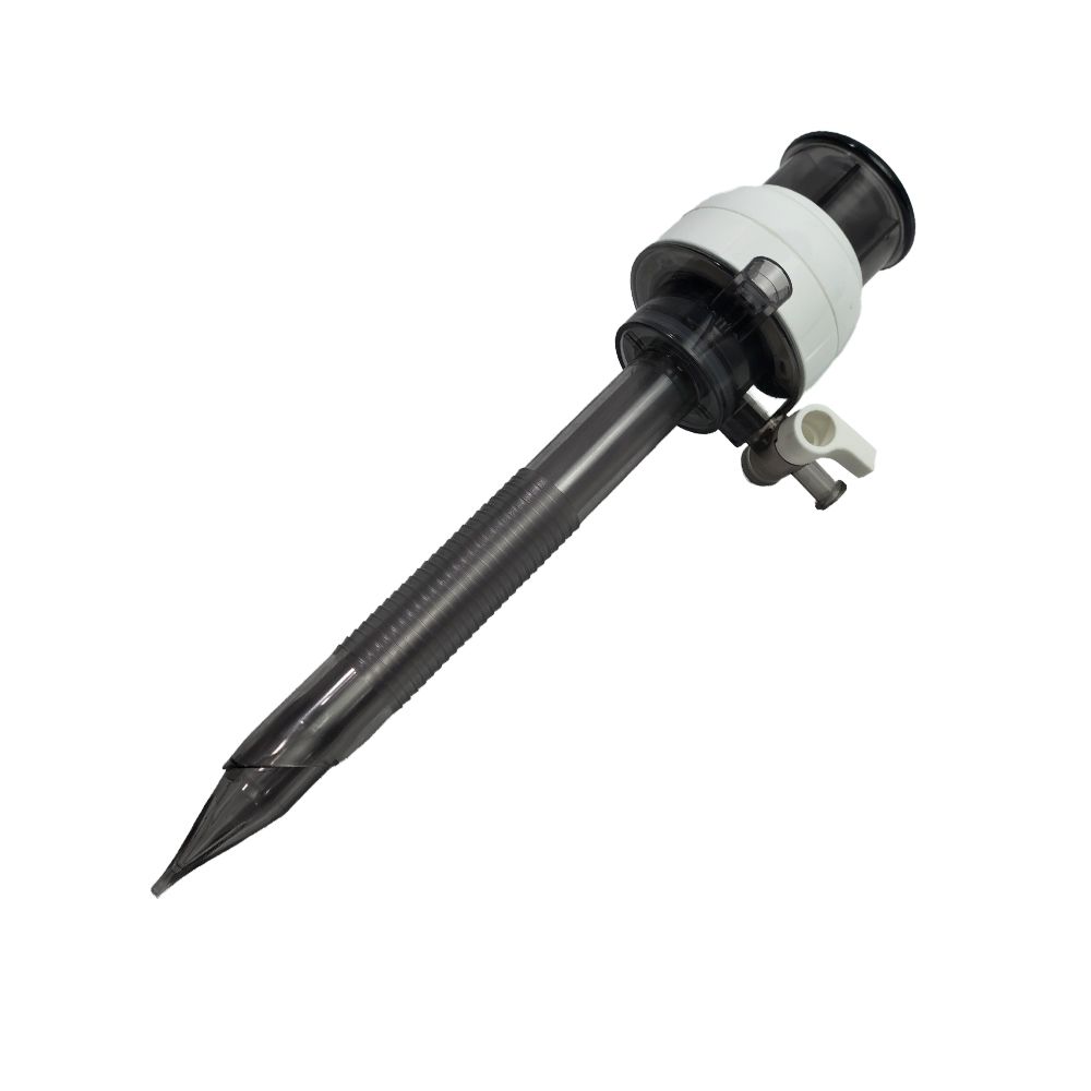
Healthcare facilities must apply incredibly rigorous standards when purchasing an optical visualization trocar. You need to balance surgeon-preferred clinical performance with objective, certifiable manufacturing metrics.
Device handle design heavily influences surgical control and precision. You must assess the grip comfort very carefully. Surgeons need a highly stable platform for their palm to exert even pressure.
The primary evaluation criteria involves managing insertion force. A device must require predictable, tightly controlled axial force. If a device requires excessive physical pressure, surgeons might push entirely too hard. This risks sudden, violent "plunging" upon finally traversing the tough fascia. A well-designed ergonomic handle prevents this dangerous overshoot. It ensures smooth, continuous entry.
Next, scrutinize the distal clear tip design meticulously. The polymer lens must be flawlessly manufactured to prevent visual artifacts.
The primary criteria here is uncompromised visibility. The tip must shield the internal laparoscope lens from heavy tissue smudging. It must also maintain absolutely zero optical distortion during the intense pressure of insertion. If the molded plastic warps the camera image, the surgeon cannot identify tissue planes accurately. They lose the primary benefit of the device.
Evaluate the universal device seal thoroughly. Quality manufacturers typically use robust duckbill or advanced multi-valve designs. These delicate internal components prevent critical gas from escaping the abdomen.
The success criteria here is exceptionally strict. The port must maintain a highly stable pneumoperitoneum at all times. Surgeons constantly swap various instruments during routine cholecystectomies. They insert thin 5mm graspers and thick 10mm clip appliers interchangeably. The dynamic seal must instantly adapt to these different diameters without leaking CO2.
Procurement teams must relentlessly enforce strict regulatory baselines. Never compromise on global manufacturing standards. You must verify current ISO 13485 certification immediately during the vendor review. Look specifically for updated FDA clearances or active CE marks. Furthermore, ensure the exclusive use of medical-grade, highly biocompatible materials. Facilities must demand entirely latex-free components to protect vulnerable patients from severe anaphylaxis.
| Evaluation Category | Key Assessment Metric | Primary Clinical Rationale |
|---|---|---|
| Ergonomic Control | Controlled axial insertion force | Prevents accidental plunging past the fascial layer into the bowel. |
| Lens Optics | Zero image distortion at the tip | Ensures highly accurate, real-time tissue plane identification. |
| Seal Integrity | Multi-valve gas retention capacity | Maintains stable pneumoperitoneum during rapid instrument changes. |
| Quality Compliance | ISO 13485 & CE/FDA clearance | Guarantees medical-grade, latex-free material safety for all patients. |
Transitioning primary entry methods requires highly intentional training. Veteran surgeons often rely heavily on tactile "clicks" when using older Veress needles. They physically feel the tissue resistance drop. When switching to modern optical ports, they must unlearn this deep tactile reliance. Instead, they must learn to interpret visual tissue compression on the monitor. It usually takes a few supervised cases to trust the screen completely. Surgical departments should plan structured proctoring for all early adopters to ensure maximum safety.
You must verify hardware compatibility before purchasing bulk hospital inventory. Different hospitals use entirely different laparoscope brands and camera models. Ensure your existing hospital laparoscope models lock securely into the newly selected vendor’s housing. Loose scopes slide backward slowly during insertion. This instantly blurs the surgeon's vital vision. Always test your specific 10mm rigid scopes in the provided sample ports before signing contracts.
Hospital administrators must conduct a highly rigorous clinical value analysis. You must carefully weigh the unit cost of disposable units against broader operational savings. Premium ports often yield hidden procedural efficiencies. They consistently facilitate faster operating room setup times. Because bladeless tips separate rather than slice tissue, patients require significantly reduced fascial closure interventions. You also completely avoid massively expensive complication-related procedures. The initial upfront device cost frequently offsets these broader surgical expenses entirely.
Never commit to a new medical vendor without extensive real-world testing. We highly recommend structuring a formal, multi-week pilot phase. Test the disposable units in the operating room during actual live procedures. Ask participating surgeons to rate the seal durability after long cases. Have them score the lens clarity under active, messy surgical conditions. Gathering this empirical data prevents incredibly costly procurement mistakes.
A brilliantly designed product means absolutely nothing if it frequently goes out of stock. Evaluate prospective vendors on their actual global production capacity. Ask for transparent, tiered volume pricing models. Scrutinize their historical lead-time stability closely. You need resilient partners who maintain robust safety stock levels domestically. Sudden supply chain interruptions halt profitable surgical schedules instantly.
Watch out carefully for common manufacturing shortcuts. We warn strictly against trocars featuring poor external thread designs. Weak or shallow threads lead directly to frustrating port back-out during active surgery. Also, firmly reject any units containing inferior internal seals. Bad seals cause a rapid, noisy loss of insufflation. This deflates the patient's abdomen rapidly and abruptly halts the entire surgical procedure.
The 10mm optical port continues to thoroughly modernize laparoscopic cholecystectomy access. It fundamentally replaces blind, dangerous tactile entry with precise, layer-by-layer visualization. The right surgical device perfectly balances clinical safety with essential operational efficiency. It enables gentle bladeless entry while fully maintaining workflow compatibility for essential gallbladder extraction tools. As modern surgical departments strive tirelessly for zero-complication outcomes, adopting superior entry tools remains a strict, non-negotiable priority.
Take the following practical steps to upgrade your surgical access protocols immediately:
Audit your current primary entry complication rates rigorously to establish a clear clinical baseline.
Form a dedicated evaluation committee comprising leading surgeons and central procurement managers.
Request detailed technical specifications and strict compliance documentation from shortlisted manufacturers.
Order clinical evaluation samples to physically test ergonomic control and seal integrity in the operating room.
A: A Hasson trocar requires a time-consuming mini-laparotomy. Surgeons manually cut through tissue layers to place the port, tying heavy sutures to secure it. It is an entirely open technique. Conversely, an optical port uses a transparent tip and an inserted laparoscope. Surgeons push it directly through the tissue, visually watching each layer on a monitor as they safely separate muscle fibers.
A: Yes. While designed primarily for initial umbilical access, surgeons occasionally use them for secondary sites. However, most surgeons prefer smaller 5mm standard ports for working sites during a cholecystectomy. Using a 10mm size for secondary sites is generally reserved for passing larger specific instruments, staplers, or addressing highly specialized extraction requirements.
A: No surgical technique completely eliminates procedural risk. However, real-time optical feedback significantly reduces the likelihood of blind puncture injuries. Surgeons can clearly see underlying adhesions or elevated bowel loops before the tip actually enters the cavity. If the surgeon applies excessive violent force or simply ignores the visual feedback on the monitor, injuries can still occur.
A: A bladeless tip gently separates fascial fibers instead of aggressively cutting them. Once the device is removed, the separated muscle fibers naturally retract and close the gap. This significantly reduces the overall size of the fascial defect. Many surgeons find they require fewer deep fascial sutures to prevent postoperative incisional hernias compared to older bladed entry.


