
Phone:
+86-139-5121-3963
Views: 0 Author: Site Editor Publish Time: 2026-07-31 Origin: Site


Establishing laparoscopic access in high-BMI patients presents unique surgical challenges. Thickened abdominal walls severely complicate primary entry. Displaced anatomical landmarks confuse even experienced surgeons. The umbilicus often shifts downwards, altering standard insertion angles. Standard access devices frequently fall short during these complex bariatric procedures. They can cause unnecessary entry trauma. They frequently lead to a frustrating loss of pneumoperitoneum. Surgeons face increased operative times when instruments slip out mid-procedure. To overcome these hurdles, surgical teams rely on advanced port technologies. An optical trocar with fixation cannula serves as a highly specialized solution. Engineers designed it specifically to handle these demanding anatomies safely. It mitigates dangerous blind entry risks by allowing real-time visualization. Furthermore, the specialized cannula design actively prevents dangerous slip-outs during high-torque movements. Implementing this device ultimately improves operative efficiency. It deeply protects patient safety. You will learn how these specialized instruments function today. We will explore their distinct clinical advantages. We will also outline the exact criteria you should evaluate when selecting them for your facility.
Visualized Entry: An optical trocar allows tissue-layer identification in real-time, reducing the risk of visceral or vascular injury during primary placement.
Stability Under Torque: The fixation cannula design actively prevents slippage and loss of pneumoperitoneum during high-torque instrument exchanges.
Optimized for Bariatrics: Extended working lengths (typically 120mm–150mm) accommodate thicker abdominal walls without compromising seal integrity.
Procurement Focus: Evaluating a bariatric trocar requires balancing per-unit cost with the reduction of operative delays and port-site complications.
Surgeons encounter immense physical hurdles when operating on patients having a high body mass index. Excess subcutaneous adipose tissue dramatically increases the distance from the skin to the peritoneal cavity. Traditional trocars often fail to bridge this deep gap effectively. They lack the necessary reach. This structural deficit forces surgeons into awkward angles. It compromises the entire surgical field. The excessive fat also exerts heavy inward pressure on the cannula.
The "slip-out" problem frequently plagues these complex operations. Bariatric instruments possess exceptionally long shafts. Manipulating these long tools creates a powerful lever effect. We call this high torque. This intense mechanical force easily dislodges standard, smooth-sleeved access ports. When you maneuver a heavy stapler, the port often pops out. This happens repeatedly in challenging cases. The thick abdominal wall acts as a rigid fulcrum. It magnifies the leverage exerted by the surgeon's hands.
Access failure introduces severe operational setbacks. When a port dislodges, you immediately lose pneumoperitoneum. This sudden deflation pauses the surgery. The abdominal dome collapses instantly. Cameras smudge as they rub against falling tissue. Surgical teams waste valuable time repositioning the port. Every minute of surgical delay increases patient risk. It prolongs anesthesia exposure needlessly. It also strains hospital resources significantly. Constant reinsertion also increases unnecessary port-site trauma. This repeated friction damages the abdominal wall. It creates localized bleeding. It severely complicates post-operative healing. You must prevent this cycle of failure.
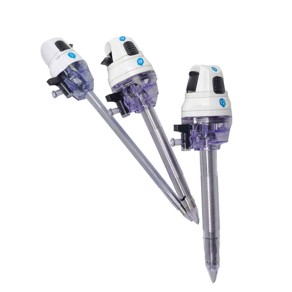
Primary entry remains a critical phase during any laparoscopic procedure. Using an optical trocar fundamentally transforms this risky step. The device accommodates a standard 0-degree laparoscope directly inside its clear obturator. This brilliant setup allows the surgeon to visualize every distinct tissue layer. You watch the high-definition monitor as the tip advances downward. You can clearly monitor the dense fascial layers. You observe the muscle fibers parting gently. Finally, you identify the thin, translucent peritoneum before piercing it safely.
Optical entry sharply contrasts traditional blind techniques. Using a standard Veress needle forces surgeons to rely strictly on tactile feedback. You wait for a subtle "pop" or a sudden loss of resistance. Blind insertion carries immense risk in high-BMI anatomies. Thick tissue severely dampens tactile sensation. Displaced organs or hidden adhesions increase the danger of inadvertent bowel injuries. Major vessel lacerations remain a catastrophic, life-threatening threat. Visualized entry mitigates these severe blind entry risks entirely. You see exactly where the instrument travels. It removes dangerous guesswork from the equation completely.
Modern devices also utilize controlled dilation to protect surrounding tissue. Bladeless optical tips separate tissue fibers rather than cutting them. They act like a wedge moving through the fascia. This blunt dissection technique creates a much smaller fascial defect. It reduces bleeding upon entry significantly. It also potentially lowers the risk of post-operative port-site hernias. Tissues naturally snap back together once you remove the cannula. This organic healing process benefits the patient immensely. It accelerates their recovery timeline.
Maintaining stable access proves just as crucial as safe entry. A robust fixation cannula guarantees operative continuity. Manufacturers engineer several unique retention designs to solve instability. Some utilize deeply threaded or ribbed outer cannulas. These aggressive threads grip the surrounding tissue firmly. Others deploy advanced balloon anchors. You inflate these small balloons once they sit inside the cavity. They pull snug against the interior wall. Some even use specialized fascial threads. These mechanisms lock the device firmly within the thick abdominal wall. They resist immense outward pulling forces.
Neutralizing torque remains the primary goal of these designs. The fixation cannula aggressively secures the laparoscopic trocar against the fascial layer. It provides a rigid, stable fulcrum. You can confidently maneuver heavy bariatric instruments without fearing displacement. You push, pull, and twist tools freely. The port stays exactly where you placed it. This stability transforms the surgical experience entirely. It reduces surgeon frustration.
Understanding how specific features drive clinical outcomes helps optimize surgical flow. You must connect mechanical design to practical utility. Here are key feature-to-outcome translations:
Feature: Advanced retention thread. Outcome: Eliminates the need for manual stabilization by the surgical assistant. They can focus on camera operation instead. It streamlines the operating room workflow.
Feature: Deep-funnel instrument entry. Outcome: Ensures smooth instrument exchanges. Tools never catch or drag on the seals. You never accidentally dislodge the cannula while pulling a stapler out.
Feature: Transparent cannula sleeve. Outcome: Allows continuous visual monitoring of the extraction. You can instantly spot hidden bleeding in the tissue tract. You address complications before the patient leaves the room.
Selecting the correct port device demands rigorous evaluation. You must match device specifications directly to patient anatomy. Working length specifications dictate success in bariatric cases. Standard lengths rarely suffice for thicker abdominal walls. Facilities usually choose between standard (100mm), bariatric (120mm), and extra-long (150mm) profiles. Your choice depends entirely on patient BMI. It also depends on the required insertion angle. A steep angle requires a significantly longer shaft.
The following comparison chart outlines working lengths and appropriate clinical applications. You should reference these guidelines during procurement.
| Profile Type | Working Length | Patient BMI Range | Optimal Insertion Application |
|---|---|---|---|
| Standard | 100mm | Below 35 | Routine access, lower angles, thinner tissue |
| Bariatric | 120mm | 35 to 45 | Thickened walls, moderate torque, standard gastric bypass |
| Extra-Long | 150mm | Above 45 | Extreme adipose layers, high torque angles, complex revisions |
Seal durability also requires close scrutiny. Bariatric operations demand dozens of instrument exchanges. You insert and remove staplers constantly. You must evaluate both primary and secondary seals. They must maintain a strict pneumoperitoneum despite continuous friction. A weak seal leaks gas rapidly. It disrupts the surgical field. It forces you to stop and reinflate the abdomen. Look for resilient duckbill seals.
Optical clarity and equipment compatibility matter equally. The distal tip must never distort the endoscope's image. Blur ruins the safety advantage of visual entry. You need a crystal-clear picture of the tissue. Ensure the port fits your facility's existing camera systems perfectly. It must accommodate 5mm or 10mm/12mm scopes flawlessly. Test them with your current equipment.
Finally, assess the required insertion force. You need a secure fixation thread. However, it must also offer an ergonomically safe insertion force. High resistance forces the surgeon to push too hard. This risks a sudden, dangerous plunge upon peritoneal entry. The device should twist smoothly into place. It should feel balanced in the hand.
Procurement teams face critical decisions when introducing new access devices. You must weigh the benefits of standardization against specialty stocking. Some hospitals standardize these advanced ports for all bariatric procedures. They use them universally. Others stock them strictly for ultra-high-BMI cases. Standardization simplifies inventory management. It reduces ordering confusion. However, it increases upfront expenditure significantly. You must balance these competing priorities.
Conducting a thorough cost-benefit analysis provides deep clarity. Specialized ports carry a premium per-unit cost. You must weigh this against the hidden costs of operating room delays. Constant slip-outs waste expensive surgical time. Every lost minute drains hospital resources. Managing preventable port-site complications also costs thousands of dollars. Investing in reliable access often yields immense procedural efficiency. You recover the premium cost through faster, safer surgeries. You avoid costly readmissions.
Facilities must also acknowledge the surgeon adoption curve. Transitioning to visualized entry requires slight technique adjustments. Deploying specific fixation mechanisms demands brief training. For example, balloon inflation protocols require practice. Proper education ensures safe and effective utilization. You should partner with vendors who provide on-site training. Support your surgical staff during this transition.
Consider taking the following sequential steps to implement these devices smoothly:
Consult with your lead bariatric surgeons. Identify current access challenges and slip-out frequencies accurately.
Request physical sample sets. Ensure they encompass 120mm and 150mm lengths for testing.
Establish a controlled evaluation period. Run this trial within a dedicated surgical review committee.
Gather concrete feedback. Focus specifically on slip-out reduction, entry smoothness, and image clarity.
Finalize a stocking protocol. Base this decision on measured clinical outcomes rather than initial price alone.
Combining visualized entry with mechanical fixation actively resolves the most persistent access failures in high-BMI surgeries. It directly tackles the dual threats of blind entry and cannula instability.
Enhance Safety Metrics: Visualized tissue tracking prevents dangerous blind organ injuries. You protect the patient at the most vulnerable stage of surgery.
Eliminate Disruptions: Deep fixation threads neutralize instrument torque. They prevent frustrating mid-surgery dislodgement entirely.
Improve Efficiency: Seamless instrument exchanges maintain stable pneumoperitoneum. This accelerates the total operative time.
These specialized ports demand a higher initial spend than standard variants. However, their clinical reliability easily justifies the investment. High-volume bariatric centers simply cannot afford preventable surgical friction. Delays compound quickly across a busy surgical schedule. We highly encourage procurement teams and surgical directors to act proactively. Request immediate sample evaluations of an advanced bariatric trocar from your suppliers. Review recent clinical literature comparing different fixation designs. Make a truly informed, evidence-based upgrade to protect your patients and empower your surgeons.
A: A specialized bariatric port typically ranges from 120mm to 150mm in working length. Standard laparoscopic ports usually measure only 100mm. The extended profile directly accommodates the thicker abdominal walls found in high-BMI patients. It guarantees the cannula reaches the peritoneal cavity securely without compromising the gas seal.
A: Yes, clinical consensus strongly supports this. A bladeless tip separates and stretches tissue fibers rather than slicing through them. This blunt dissection creates a significantly smaller fascial defect. Once the cannula is removed, the separated muscle layers naturally contract. This dramatically reduces the risk of post-operative herniation.
A: Extraction depends entirely on the specific retention design. You can remove a threaded cannula rapidly by simply unthreading it with a firm counter-clockwise twist. Balloon-anchored variations require you to deflate the retaining balloon first. Deflation usually takes merely a few seconds via a quick-release valve.
A: The vast majority of these highly engineered devices are disposable and single-use. While hybrid options exist, a single-use model guarantees flawless optical clarity for the laparoscope. It also ensures the delicate primary and secondary gas seals remain pristine. Reusing these complex mechanisms often compromises crucial patient safety.


